Ultrasound-guided wrist arthrocentesis

wrist+aspiration+pic+001.jpg


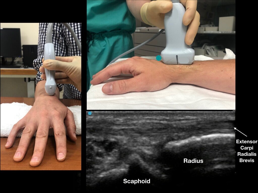
Figure 1a - Dorsal aspect of the distal forearm and radiocarpal joint. Note Lister’s tubercle and its relation to the scaphoid bone.

Figure 1b - A roll is placed on the palmar aspect of the wrist for slight flexion. With the ultrasound probe in a transverse orientation, slide distally until Lister’s tubercle is visualized. The extensor carpi radialis brevis tendon (ECRB) and extensor carpi radialis longus (ECRL) can be seen on the radial aspect of Lister’s tubercle.

wrist+aspiration+pics.002.jpg

Figure 2 - Rotate the ultrasound transducer in a longitudinal plane (as compared to the radius) with the probe marker pointing distal. In a patient with no effusion, the distal radius and scaphoid bone with minimal anechoic fluid should be noted.

wrist aspiration pics.003.jpeg

Figure 3 - In a patient with a radoiocarpal effusion, note the anechoic (black) fluid just anterior to the scaphoid. The extensor carpi radials brevis acts as a good ultrasound landmark.

wrist aspiration pics.004.jpeg

Figure 4 - With the ultrasound probe covering the extensor carpi radials brevis, enter the swollen joint capsule with an out-of-plane technique. We recommend a steep needle angle of entry between Lister Tubercle and extensor carpi radials brevis. The procedure should be performed with sterile precautions (like any other joint aspiration).