

superficial cervical plexus block © 2012 by Arun Nagdev is licensed under CC BY-NC-ND 4.0

A. The distribution of the SCP block. Note that the innervation extends down to the T2 level and to the cape of the shoulder.
B. Common emergency medicine indications for the SCP block
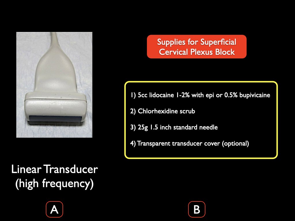
A. A high-frequency linear transducer is ideal for the ultrasound-guided SCP block.
B. Supplies needed for the SCP block.

A. Place the transducer in a transverse orientation to the neck at the superior portion of the thyroid cartilage. Note the classic ultrasonographic landmarks; CA= carotid artery; IJ= internal jugular vein; SCM= sternocleidomastiod muscle
B. At the level of the superior portion of the thyroid cartilage, slowly slide the transducer laterally. Note the tapering of the SCM muscle and the levator scapulae muscle (LSM) just below. The superior cervical plexus (SCP- denoted by the blue arrowheads) is the group of hyperechoic structures between the SCM muscle and LCM.

A. The ultrasound screen is contralateral to the affected extremity with a clear view of the screen for the clinician.
B. The needle and needle tip (red arrows) are visualized clearly entering from the lateral aspect of the ultrasound image. Note that the ultrasound transducer directional marker is medial (yellow arrow). Anechoic anesthetic is being placed in between the SCM muscle and LSM, with clear visualization of the needle tip as well as the SCP (blue arrowheads).

Superficial cervical plexus block.
Most find this an EZ block. Very satisfying for:
1. Clavicle fractures
2. I&D of those abscess patients get under the chin in the shaving area.
3. Lacerations of the ear lobe are blocked as well.
4. IJ central lines. Good practice and if they are awake, really makes it a completely painless procedure.
Print out a PDF of our self-guided
workbook
Additionally references are available as PDFS here & here.
A brief video from our friends at Nagoya University is below along with brief curriculum.
