

A simplified technique for ultrasound-guided pericardiocentesis.
Download the pdf here
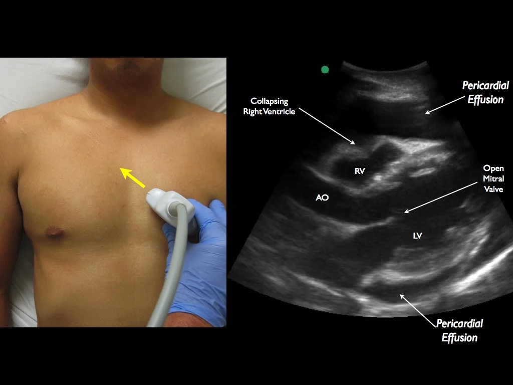
1) Curvilinear Probe
2) Probe marker to the patient’s right shoulder
3) Adjust probe to find largest pocket of fluid

**Tamponade is a clinical diagnosis. A plethoric IVC may assist in determining elevated right sided pressures.
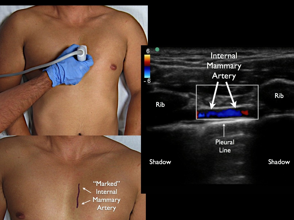
1) If effusion very midline, locate the mammary arteries with Linear Probe (optional)

1) Ultrasound screen in field of view
2) Local anesthetic in skin and muscle
3) Low dose etomidate or ketamine just before entering pericardial sac
4) In-plane with needle visualization

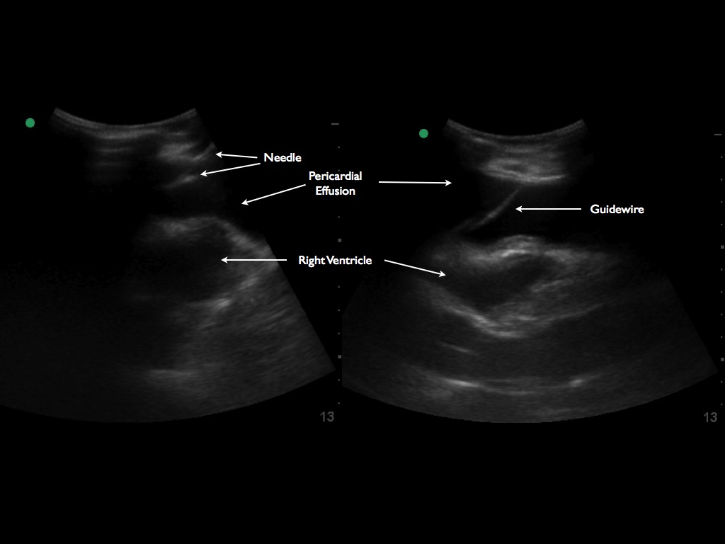
1) Visualize the guidewire to confirm placement

1) Drain effusion
2) Confirm reduction in pericardial effusion and IVC collapse